
Histopathology Biopsy: Large Complex / Cancer Specimen Test
Short Name: Large Complex Biopsy
Also known as: Large Tissue Biopsy Histopathology, Cancer Specimen Histopathology, Surgical Pathology - Large Specimen, Oncology Resection Histopathology
Histopathology Biopsy: Large Complex / Cancer Specimen Test test available at DNA Labs India for ₹6,000. Uses Tissue Processing, Paraffin Embedding, Microtomy, H&E Staining, Special Stains (as indicated), Microscopy by Pathologist on Surgical tissue specimen in 10% Formal-saline samples. Results in Sample accepted daily by 6 PM. Standard report delivery: 9 working days. For bone specimens: 16–18 working days.. Free home collection in 300+ cities across India.

🩺 Medically Reviewed By
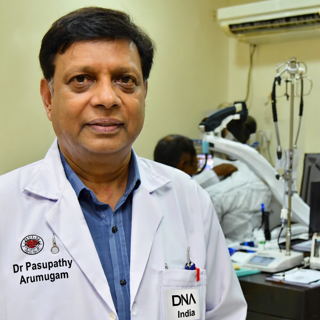
Dr Pasupathy Arumugam
Consultant Pathologist · Reg: 21521
Last reviewed: September 7, 2026
Overview
This test is performed on large surgical resection specimens to confirm a cancer diagnosis, determine tumour type, grade, and stage, evaluate surgical margin status, detect lymphovascular or perineural invasion, and identify features that guide further treatment. Common specimen types include: esophagectomy, gastrectomy, mastectomy, hemi/total colectomy, large bone resection, ovarian tumour resection, radical nephrectomy for cancer, radical neck dissection, radical hysterectomy, radical orchidectomy, soft tissue tumour resections, and head and neck resections.
- Test Code
- 763
- CPT Code
- 88305
- ICD Code
- C80.1
- Price
- ₹6,000
- Sample Type
- Surgical tissue specimen in 10% Formal-saline
- Result Time
- Sample accepted daily by 6 PM. Standard report delivery: 9 working days. For bone specimens: 16–18 working days.
- Fasting Required
- No
- Method
- Tissue Processing, Paraffin Embedding, Microtomy, H&E Staining, Special Stains (as indicated), Microscopy by Pathologist
Sample Collection
The treating surgeon or oncologist must complete the Surgical Pathology Requisition Form for Oncology Resections (Form 3) with complete brief clinical history, prior treatment details, and provisional diagnosis. For patients with multiple specimens, all specimens must be registered under a single lab number.
Method: Surgical resection / excision by treating surgeon or oncologist
Laboratory Analysis
The surgical specimen is excised by the treating surgeon as part of the planned oncological procedure. The specimen must be immediately placed in a leak-proof container with 10% formal-saline ensuring the specimen is completely submerged.
Report Delivery
The container is labelled with patient details and transported to the laboratory at room temperature. The specimen undergoes grossing, processing, embedding, sectioning, staining, and microscopic examination by a qualified pathologist.
Timeline: Sample accepted daily by 6 PM. Standard report delivery: 9 working days. For bone specimens: 16–18 working days.
Patient Instructions
About This Test
Who Should Get This Test
This test is performed on large surgical resection specimens to confirm a cancer diagnosis, determine tumour type, grade, and stage, evaluate surgical margin status, detect lymphovascular or perineural invasion, and identify features that guide further treatment. Common specimen types include: esophagectomy, gastrectomy, mastectomy, hemi/total colectomy, large bone resection, ovarian tumour resection, radical nephrectomy for cancer, radical neck dissection, radical hysterectomy, radical orchidectomy, soft tissue tumour resections, and head and neck resections.
How to Prepare
- Submit the surgical specimen in 10% formal-saline in a leak-proof container
- Formal-saline must completely cover the specimen
- Ship the specimen at room temperature
- Complete the Surgical Pathology Requisition Form for Oncology Resections (Form 3) with brief clinical history — this is mandatory
- For patients with multiple specimens, register all specimens under a single lab number
- Label the container clearly with patient name, lab number, and specimen site
Doctor's Notes
Reviewed by Dr Pasupathy Arumugam — MBBS, MD (Pathology) · Reg. No. 21521
"Histopathological examination of large surgical resection specimens is the gold standard for confirming a cancer diagnosis, determining tumor type, grade, margin status, and staging. Accurate pathology reporting directly influences treatment decisions including chemotherapy, radiation, or further surgery. Patients should ensure complete clinical history and prior treatment details accompany the specimen for optimal interpretation."
Last medically reviewed: September 7, 2026
Test Parameters & Specifications
Sample Stability
- Specimen received without 10% formal-saline or improperly fixed
- Specimen container leaking or damaged in transit
- Surgical Pathology Requisition Form (Form 3) not submitted or incomplete clinical history
- Specimen not labelled or patient identifiers mismatched
- Specimen received in a completely dried state with no fixative
Understanding Your Results
Negative margins (clear margins)
No cancer cells detected at the surgical edges, suggesting complete excision of the tumour.
Positive margins
Cancer cells present at one or more surgical edges, indicating residual disease may remain. Further surgery or adjuvant therapy may be recommended.
High-grade tumour
Poorly differentiated or undifferentiated cells indicating aggressive behaviour and potentially requiring more intensive treatment.
Lymphovascular invasion present
Cancer cells detected in blood or lymphatic vessels, suggesting higher risk of metastasis.
Positive lymph nodes
Metastatic cancer found in regional lymph nodes, which influences the pathological stage and may necessitate systemic therapy.
Complete pathological response (in post-neoadjuvant specimens)
No viable tumour cells found in the resected specimen after pre-operative chemotherapy or radiation, indicating excellent treatment response.
Consult your oncologist or treating physician to discuss the histopathology report findings, cancer stage, and recommended treatment plan. If the report indicates positive margins, high-grade tumour, lymphovascular invasion, or positive lymph nodes, prompt follow-up is essential for planning further treatment.
Limitations
- ⚠Histopathological examination is limited to the tissue submitted; it may not represent the entire tumour if sampling is incomplete
- ⚠Certain well-differentiated tumours may require immunohistochemistry (IHC) or molecular testing for definitive classification, which may incur additional cost and turnaround time
- ⚠Small foci of metastasis in lymph nodes may occasionally be missed on routine H&E staining
- ⚠Post-treatment specimens may show extensive necrosis making accurate grading difficult
- ⚠This test evaluates morphology only; molecular or genetic profiling of the tumour requires separate testing
Risks & Considerations
- ●This test involves examination of an already resected surgical specimen; there are no additional risks from the histopathology procedure itself
- ●Risks associated with the surgical procedure to obtain the specimen (bleeding, infection, anaesthesia-related risks) are managed by the surgical team
- ●Delayed fixation may compromise tissue quality and affect diagnostic accuracy
Interfering Factors
- ●Inadequate fixation — specimen not fully covered in 10% formal-saline may show autolysis artefacts
- ●Excessive delay in placing specimen in fixative after surgical removal
- ●Incompletely labelled specimen or mismatched requisition form
- ●Absence of detailed clinical history on the Surgical Pathology Requisition Form (Form 3)
- ●Previous radiation or chemotherapy changes may complicate histological interpretation
Compare With Similar Tests
| Test | Histopathology Biopsy: Large Complex / Cancer Specimen Test | Fine Needle Aspiration Cytology (FNAC) | Core Needle Biopsy | Immunohistochemistry (IHC) | Frozen Section Biopsy |
|---|---|---|---|---|---|
| Comparison | Histopathology Biopsy: Large Complex / Cancer Specimen Test | FNAC uses a thin needle to aspirate cells for cytological evaluation. It is minimally invasive but provides limited tissue architecture. Large complex specimen histopathology provides full tissue architecture for definitive diagnosis and staging. | Core needle biopsy retrieves a small cylinder of tissue. It is useful for initial diagnosis but does not provide information about margins or full tumour extent. Large specimen histopathology evaluates the entire resected tumour. | IHC uses antibodies to detect specific proteins in tissue sections. It is often performed as a supplementary test after routine histopathology to subtype tumours or guide targeted therapy decisions. | Frozen section provides rapid intra-operative diagnosis within minutes but with lower image quality. Formalin-fixed histopathology provides superior morphological detail and is the gold standard for final diagnosis. |
Frequently Asked Questions
What is a histopathology biopsy of a large complex or cancer specimen?
How much does the Histopathology Biopsy: Large Complex / Cancer Specimen Test cost at DNA Labs India?
What types of specimens are included in this test?
How should the specimen be submitted?
Is fasting required before this test?
How long does it take to get the report?
Is home sample collection available for this test?
What information does the histopathology report contain?
What does a positive margin mean in the report?
Can this test detect all types of cancer?
Do I need a doctor's prescription for this test?
Is this test covered under government health schemes like PMJAY or CGHS?
Related Tests
Reference Laboratory Services
We serve as a reference laboratory for hospitals and clinics across India. Send samples from your facility with same-day pickup, priority processing, and results delivered through our online portal. Competitive institutional pricing available.
Your Data Privacy
Your medical data is protected under Indian law.
✓ Stored in India: All patient records are stored on servers located in India. No data is transferred outside the country.
✓ DPDP Act Compliant: Under the Digital Personal Data Protection Act 2023, you can request deletion of your records at any time by contacting support.
Book Your Test
Enter your details and we'll connect you within 15 minutes.