
Immunohistochemistry CD4 Test
Short Name: IHC CD4
Also known as: CD4 Immunostain, CD4 IHC Test, IHC CD4 Marker, CD4 Tissue Staining, Immunohistochemistry CD4 Panel
Immunohistochemistry CD4 Test test available at DNA Labs India for ₹3,000. Uses Immunohistochemistry (IHC), Manual and automated tissue staining, Light microscopy evaluation on Tumor tissue in 10% Formal-saline or Formalin Fixed Paraffin Embedded (FFPE) block samples. Results in Sample Daily by 6 pm; Reports available in 5 business days for tissue blocks and biopsies, and 7 business days for large complex tissue specimens.. Free home collection in 300+ cities across India.

🩺 Medically Reviewed By
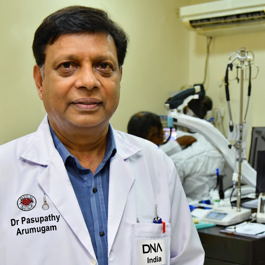
Dr Pasupathy Arumugam
Consultant Pathologist · Reg: 21521
Last reviewed: September 7, 2026
Overview
The Immunohistochemistry CD4 Test is performed to identify the expression of the CD4 antigen on tissue sections. This serves several key diagnostic purposes: it aids in the classification of T-cell lymphomas such as Mycosis Fungoides and Sézary syndrome; it supports the determination of T-cell or histiocytic lineage in hematolymphoid neoplasms; and it helps pathologists differentiate between various subtypes of lymphoproliferative disorders. CD4 staining, when combined with other immunohistochemical markers such as CD3, CD8, CD20, and CD30, provides a comprehensive immunophenotypic profile that is essential for accurate diagnosis, prognostication, and treatment planning in oncology.
- Test Code
- 975
- CPT Code
- 88342
- ICD Code
- C84
- Price
- ₹3,000
- Sample Type
- Tumor tissue in 10% Formal-saline or Formalin Fixed Paraffin Embedded (FFPE) block
- Result Time
- Sample Daily by 6 pm; Reports available in 5 business days for tissue blocks and biopsies, and 7 business days for large complex tissue specimens.
- Fasting Required
- No
- Method
- Immunohistochemistry (IHC), Manual and automated tissue staining, Light microscopy evaluation
Sample Collection
Ensure a copy of the histopathology report, site of biopsy, and detailed clinical history are available to accompany the specimen. The tissue should be properly fixed in 10% Formal-saline or submitted as a Formalin Fixed Paraffin Embedded (FFPE) block.
Method: Tissue biopsy / Surgical excision
Laboratory Analysis
Tissue is collected via biopsy or surgical excision by the treating surgeon or oncologist. The specimen is immediately placed in 10% Formal-saline or processed into an FFPE block as per standard histopathology protocols.
Report Delivery
Ship the specimen at room temperature along with a copy of the histopathology report, site of biopsy, and clinical history. No special post-collection care is required for the patient as the tissue has already been excised.
Timeline: Sample Daily by 6 pm; Reports available in 5 business days for tissue blocks and biopsies, and 7 business days for large complex tissue specimens.
Patient Instructions
About This Test
Who Should Get This Test
The Immunohistochemistry CD4 Test is performed to identify the expression of the CD4 antigen on tissue sections. This serves several key diagnostic purposes: it aids in the classification of T-cell lymphomas such as Mycosis Fungoides and Sézary syndrome; it supports the determination of T-cell or histiocytic lineage in hematolymphoid neoplasms; and it helps pathologists differentiate between various subtypes of lymphoproliferative disorders. CD4 staining, when combined with other immunohistochemical markers such as CD3, CD8, CD20, and CD30, provides a comprehensive immunophenotypic profile that is essential for accurate diagnosis, prognostication, and treatment planning in oncology.
How to Prepare
- Submit tumor tissue in 10% Formal-saline or as a Formalin Fixed Paraffin Embedded (FFPE) block
- Ship the specimen at room temperature
- Provide a clear copy of the Histopathology report with the sample
- Mention the exact site of biopsy on the requisition form
- Include detailed clinical history and provisional diagnosis
- Ensure the container is properly labeled with patient details and sample ID
Doctor's Notes
Reviewed by Dr Pasupathy Arumugam — MBBS, MD (Pathology) · Reg. No. 21521
"Immunohistochemistry for CD4 is an invaluable adjunct in the diagnostic workup of cutaneous T-cell lymphomas such as Mycosis Fungoides and Sézary syndrome. It helps establish T-cell or histiocytic lineage in hematolymphoid neoplasms when morphological assessment alone is inconclusive. Clinicians should correlate IHC CD4 results with the complete immunohistochemical panel, clinical findings, and histopathology report for accurate diagnosis and staging."
Last medically reviewed: September 7, 2026
Test Parameters & Specifications
Sample Stability
- Unlabeled or mislabeled specimen
- Specimen received without histopathology report or clinical history
- Tissue fixed in incorrect fixative or improperly processed
- Severely autolyzed or necrotic tissue with no viable diagnostic material
- Specimen shipped at inappropriate temperature or damaged during transit
Understanding Your Results
CD4 Positive (strong membranous staining)
Supports T-helper cell or histiocytic lineage. In the context of cutaneous lymphoproliferative lesions, strong CD4 positivity is consistent with Mycosis Fungoides or Sézary syndrome. Correlation with clinical findings and other IHC markers is essential.
CD4 Positive (weak to moderate staining)
May indicate partial CD4 expression seen in some T-cell lymphomas or reactive conditions. Further workup with additional IHC markers and clinical correlation is recommended.
CD4 Negative
Absence of CD4 expression may indicate a CD8-positive T-cell lymphoma, NK-cell lymphoma, B-cell lymphoma, or a non-lymphoid neoplasm. Additional markers such as CD3, CD8, CD20, CD56, and TCR gene rearrangement studies may be warranted.
Equivocal / Non-contributory
Insufficient tissue, poor fixation, or technical issues may result in equivocal results. Repeat testing on a better-quality specimen or alternative diagnostic methods such as flow cytometry may be considered.
Consult your oncologist or pathologist if your IHC CD4 result is positive, negative but clinical suspicion persists, or if the result is equivocal. Any new or worsening skin lesions, lymphadenopathy, unexplained weight loss, or recurring infections should prompt immediate medical evaluation. Your doctor will integrate the IHC CD4 findings with the full immunohistochemical panel, clinical history, and imaging studies to arrive at a definitive diagnosis and treatment plan.
Limitations
- ⚠This test is qualitative and semi-quantitative; it does not provide absolute cell counts
- ⚠Results must be interpreted in conjunction with morphological findings and a full immunohistochemical panel
- ⚠CD4 is not exclusive to T-helper cells and may be expressed on monocytes, macrophages, and dendritic cells
- ⚠Antigen retrieval efficacy may vary between laboratories and tissue processing methods
- ⚠Cannot replace flow cytometry for detailed immunophenotyping in all clinical scenarios
Risks & Considerations
- ●No direct risks to the patient as the test is performed on an already excised tissue specimen
- ●There is a small possibility of false-negative or equivocal results requiring repeat testing
- ●Misinterpretation without correlation to full clinical context may lead to incorrect diagnosis
Interfering Factors
- ●Inadequate tissue fixation may lead to false-negative results
- ●Prolonged fixation in formalin beyond recommended duration can degrade antigen epitopes
- ●Decalcification agents may adversely affect CD4 antigen detection
- ●Necrotic or heavily inflamed tissue may yield ambiguous staining patterns
- ●Use of non-standard fixation protocols at the referring facility
Compare With Similar Tests
| Test | Immunohistochemistry CD4 Test | Flow Cytometry CD4 Panel | CD4 Blood Count (HIV Monitoring) | Immunohistochemistry CD3 Test |
|---|---|---|---|---|
| Comparison | Immunohistochemistry CD4 Test | Flow cytometry provides quantitative CD4 cell counts from blood or tissue suspensions, while IHC CD4 evaluates CD4 expression in intact tissue architecture. IHC is preferred for solid tissue evaluation, whereas flow cytometry is better suited for blood and bone marrow samples. | CD4 blood count measures circulating CD4+ T-helper cell numbers in peripheral blood, primarily used for HIV monitoring. IHC CD4 detects CD4 protein expression in tissue sections for cancer diagnosis. These are fundamentally different tests serving different clinical purposes. | CD3 is a pan T-cell marker expressed on all T-cells, while CD4 is expressed specifically on the helper T-cell subset. CD3 is often used alongside CD4 to confirm T-cell lineage, whereas CD4 helps subtype the T-cell population. |
Frequently Asked Questions
What is the Immunohistochemistry CD4 Test?
What is the cost of the Immunohistochemistry CD4 Test in India?
What sample is required for the IHC CD4 Test?
How long does it take to get the IHC CD4 Test results?
Is fasting required for the Immunohistochemistry CD4 Test?
Who should undergo the Immunohistochemistry CD4 Test?
What does a positive CD4 result mean?
What does a negative CD4 result mean?
Is the Immunohistochemistry CD4 Test the same as a CD4 blood count?
Is home sample collection available for the IHC CD4 Test?
What other tests are commonly ordered along with the IHC CD4 Test?
How should I prepare for the Immunohistochemistry CD4 Test?
Related Tests
Reference Laboratory Services
We serve as a reference laboratory for hospitals and clinics across India. Send samples from your facility with same-day pickup, priority processing, and results delivered through our online portal. Competitive institutional pricing available.
Your Data Privacy
Your medical data is protected under Indian law.
✓ Stored in India: All patient records are stored on servers located in India. No data is transferred outside the country.
✓ DPDP Act Compliant: Under the Digital Personal Data Protection Act 2023, you can request deletion of your records at any time by contacting support.
Book Your Test
Enter your details and we'll connect you within 15 minutes.