
Immunohistochemistry Thyroid Carcinoma Panel Test
Short Name: IHC Thyroid Carcinoma Panel
Also known as: IHC Thyroid Panel, Thyroid Carcinoma IHC Panel, Thyroid Cancer Immunohistochemistry Panel, IHC Panel for Thyroid Tumor Subtyping
Immunohistochemistry Thyroid Carcinoma Panel Test test available at DNA Labs India for ₹5,265. Uses Immunohistochemistry (IHC) on Tumor tissue in 10% Formal-saline OR Formalin Fixed Paraffin Embedded (FFPE) block samples. Results in Tissue biopsy specimens: 5 working days. Large complex tissue specimens: 7 working days. Reports available daily by 6 PM on completion.. Free home collection in 300+ cities across India.

🩺 Medically Reviewed By
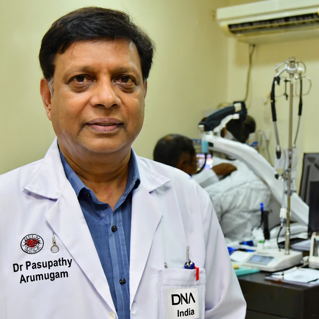
Dr Pasupathy Arumugam
Consultant Pathologist · Reg: 21521
Last reviewed: September 7, 2026
Overview
The Immunohistochemistry Thyroid Carcinoma Panel Test is ordered to confirm the diagnosis of thyroid carcinoma, differentiate between histological subtypes of thyroid cancer (papillary, follicular, medullary, anaplastic), identify markers that guide targeted therapy decisions, and help determine the origin of metastatic tumours suspected to be of thyroid origin. It is an essential tool for oncologists and pathologists in establishing a definitive diagnosis and planning treatment.
- Test Code
- 1057
- ICD Code
- C73
- Price
- ₹5,265
- Sample Type
- Tumor tissue in 10% Formal-saline OR Formalin Fixed Paraffin Embedded (FFPE) block
- Result Time
- Tissue biopsy specimens: 5 working days. Large complex tissue specimens: 7 working days. Reports available daily by 6 PM on completion.
- Fasting Required
- No
- Method
- Immunohistochemistry (IHC)
Sample Collection
Provide a copy of the histopathology report, site of biopsy, and clinical history along with the tissue specimen. No special patient preparation such as fasting is required for tissue submission.
Method: Tissue biopsy / Surgical resection specimen
Laboratory Analysis
Tissue is collected via biopsy (FNAB, core needle biopsy) or surgical excision by the treating physician. The specimen is immediately placed in 10% Formal-saline or submitted as a formalin-fixed paraffin-embedded (FFPE) block.
Report Delivery
Specimen is labelled accurately and shipped at room temperature to the laboratory. A copy of the histopathology report, biopsy site, and clinical history must accompany the specimen.
Timeline: Tissue biopsy specimens: 5 working days. Large complex tissue specimens: 7 working days. Reports available daily by 6 PM on completion.
Patient Instructions
About This Test
Who Should Get This Test
The Immunohistochemistry Thyroid Carcinoma Panel Test is ordered to confirm the diagnosis of thyroid carcinoma, differentiate between histological subtypes of thyroid cancer (papillary, follicular, medullary, anaplastic), identify markers that guide targeted therapy decisions, and help determine the origin of metastatic tumours suspected to be of thyroid origin. It is an essential tool for oncologists and pathologists in establishing a definitive diagnosis and planning treatment.
How to Prepare
- Submit tumour tissue in 10% Formal-saline OR Formalin Fixed Paraffin Embedded (FFPE) block.
- Ship the specimen at room temperature.
- Provide a copy of the histopathology report with the specimen.
- Mention the site of biopsy clearly on the requisition form.
- Include relevant clinical history including prior treatment details.
Doctor's Notes
Reviewed by Dr Pasupathy Arumugam — MBBS, MD (Pathology) · Reg. No. 21521
"The Immunohistochemistry Thyroid Carcinoma Panel is a critical diagnostic tool for confirming and subtyping thyroid malignancies. By evaluating a combination of markers including thyroglobulin, calcitonin, CK-19, HBME-1, BRAF, synaptophysin, and PAX-8, this panel helps distinguish between papillary, follicular, medullary, and anaplastic thyroid carcinomas. Accurate subtyping directly influences treatment planning, surgical approach, and prognosis. I recommend this panel for any case where the histopathological diagnosis of thyroid carcinoma needs immunohistochemical confirmation or where the subtype is uncertain on routine H&E staining."
Last medically reviewed: September 7, 2026
Test Parameters & Specifications
Sample Stability
- Specimen received without proper fixation or in dried condition
- No accompanying histopathology report or clinical history
- Specimen with extensive necrosis or autolysis making evaluation impossible
- Specimen container damaged or leaking
- Unlabelled or mislabelled specimen
Understanding Your Results
Findings are consistent with Papillary Thyroid Carcinoma (PTC). BRAF V600E positivity may indicate a more aggressive subtype.
Findings may suggest Follicular Thyroid Carcinoma (FTC). Clinical correlation and histological assessment of capsular/vascular invasion are required.
Findings are characteristic of Medullary Thyroid Carcinoma (MTC), a tumour of parafollicular C-cells. Serum calcitonin and CEA levels should be correlated.
May suggest Anaplastic (Undifferentiated) Thyroid Carcinoma or a non-thyroidal malignancy. Additional workup including PAX-8 and clinical correlation is recommended.
PAX-8 positivity confirms thyroid origin of the tumour, which is particularly useful when evaluating metastatic disease of unknown primary.
Consult your oncologist or endocrinologist if you have a thyroid nodule, abnormal thyroid ultrasound findings, unexplained neck swelling, or if your treating physician has recommended this panel for further evaluation of a thyroid tissue biopsy. Early and accurate diagnosis is critical for effective treatment of thyroid carcinoma.
Limitations
- ⚠IHC results should always be interpreted in conjunction with clinical findings, imaging, and routine histopathology (H&E staining).
- ⚠No single marker is 100% specific for any thyroid carcinoma subtype; a panel approach is recommended.
- ⚠Rare or poorly differentiated thyroid carcinomas may show atypical staining patterns.
- ⚠This test does not replace molecular genetic testing when indicated.
Risks & Considerations
- ●This test is performed on an already collected tissue specimen; there are no additional risks to the patient from the IHC procedure itself.
- ●Risks associated with the original biopsy or surgical procedure (bleeding, infection, scarring) should be discussed with the treating physician.
Interfering Factors
- ●Inadequate or poorly preserved tissue specimen
- ●Prolonged ischemia time before tissue fixation
- ●Over-fixation or under-fixation in formalin
- ●Decalcification of tissue may affect antigen integrity
- ●Previous treatment with chemotherapy or radiation may alter marker expression
Compare With Similar Tests
| Test | Immunohistochemistry Thyroid Carcinoma Panel Test | Thyroid Profile (T3, T4, TSH) | Fine Needle Aspiration Cytology (FNAC) | Thyroid Ultrasound | Thyroglobulin Blood Test |
|---|---|---|---|---|---|
| Comparison | Immunohistochemistry Thyroid Carcinoma Panel Test | Thyroid function tests evaluate hormonal status of the thyroid gland but cannot detect or subtype thyroid cancer. The IHC panel is used specifically for cancer diagnosis and subtyping on tissue specimens. | FNAC provides an initial cytological assessment of thyroid nodules and may be indeterminate (Bethesda III/IV categories). The IHC panel is used when tissue biopsy is available to provide a definitive immunohistochemical diagnosis. | Ultrasound is a first-line imaging tool for evaluating thyroid nodules but cannot confirm malignancy or subtype. The IHC panel provides definitive tissue-level diagnosis. | Serum thyroglobulin is used as a tumour marker for monitoring differentiated thyroid carcinoma after treatment. The IHC tissue-based thyroglobulin staining confirms follicular cell origin of the tumour. |
Frequently Asked Questions
What is the Immunohistochemistry Thyroid Carcinoma Panel Test?
What markers are included in this IHC panel?
Who should get this test done?
What sample is required for this test?
Is fasting required for this test?
What is the cost of the Immunohistochemistry Thyroid Carcinoma Panel Test?
How long does it take to get the results?
What does a positive CK-19 and HBME-1 result mean?
Can this test detect medullary thyroid carcinoma?
Is the IHC Thyroid Panel Test available in my city?
Is this test covered under government health schemes like PMJAY or CGHS?
Can I use an old tissue block or previously collected biopsy for this test?
Related Tests
Reference Laboratory Services
We serve as a reference laboratory for hospitals and clinics across India. Send samples from your facility with same-day pickup, priority processing, and results delivered through our online portal. Competitive institutional pricing available.
Your Data Privacy
Your medical data is protected under Indian law.
✓ Stored in India: All patient records are stored on servers located in India. No data is transferred outside the country.
✓ DPDP Act Compliant: Under the Digital Personal Data Protection Act 2023, you can request deletion of your records at any time by contacting support.
Book Your Test
Enter your details and we'll connect you within 15 minutes.